SuperTeacherTools SuperTeacherTools Help
Jeopardy Home Create a New Game Create a New Game
How to Use Instant Jeopardy Review:

Instant Jeopardy Review is designed for live play with up to ten individuals or teams. Teams choose a question, then try to give the best answer. Scoring is built in for each team. You can post a link to this review game using the orange game information button below. If you are the creator of this game, you can edit the game with the red edit button. Have fun!


Seconds To Answer Each Question
Set to 0 to hide the timer

male

Anatomy Ultrasound CT & MRI General Pandora's box
10 10 10 10 10
20 20 20 20 20
30 30 30 30 30
40 40 40 40 40
50 50 50 50 50
Final Question
Edit Game

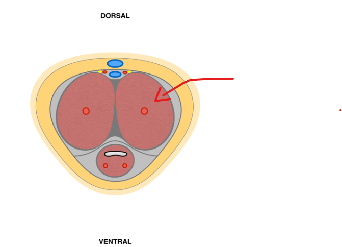
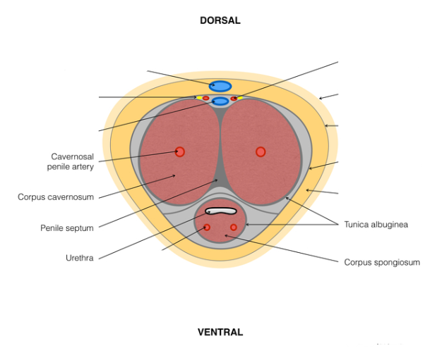
what is the name of the following structure.

View Answer

what is the name of the following structure.


corpus cavernosum


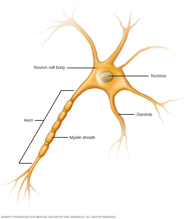
Innervation of the Male phallus.Nerve cell (neuron) - Mayo Clinic

View Answer

Innervation of the Male phallus.Nerve cell (neuron) - Mayo Clinic


spinal cord segments S2 - S4.

cavernous, pudendal, and dorsal nerves of the penis. (2 at least)Penile innervation. Adapted from Brackett et al. Treatment of... | Download  Scientific Diagram


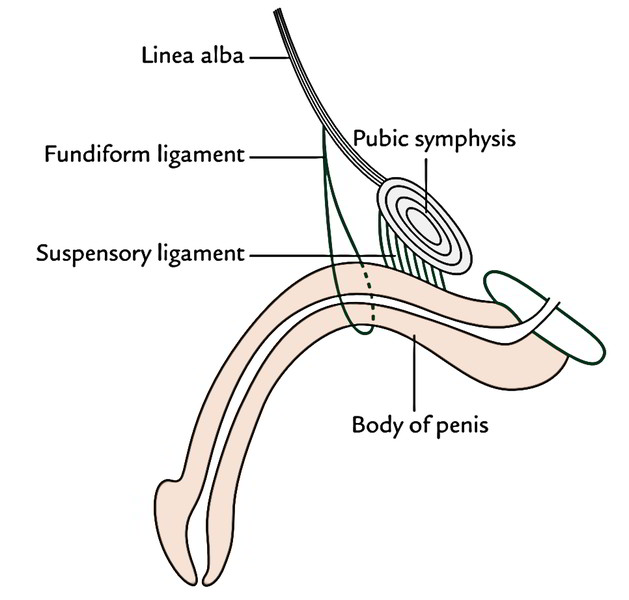
The root of the penis is supported by two ligaments, name them.Gym Memes - Anyone else?! 😅 | Facebook

View Answer

The root of the penis is supported by two ligaments, name them.Gym Memes - Anyone else?! 😅 | Facebook


Suspensory ligament

Fundiform ligamentPenis Anatomy And Physiology – Earth's Lab


name the anatomical anomaly that results from failure of the urethral folds to close.

View Answer

name the anatomical anomaly that results from failure of the urethral folds to close.


Hypospadias

Hypospadias - Classification - Management - TeachMePaeds


vasculature of the prostate and its innervation.

View Answer

vasculature of the prostate and its innervation.


prostatic arteries

prostatic venous plexus

inferior hypogastric plexus


name the anatomical structure.

View Answer

name the anatomical structure.


prostate


name the 2 anatomical structures.

View Answer

name the 2 anatomical structures.


testes and the epididymis.


what commonly calcifies in the prostate with age?

View Answer

what commonly calcifies in the prostate with age?


calcification of the prostatic secretion forming corpora amylacea.


what is the primary differential for the following:

https://radiopaedia.org/cases/75784/studies/87155?lang=gb

View Answer

what is the primary differential for the following:

https://radiopaedia.org/cases/75784/studies/87155?lang=gb


Seminoma


U/S:

  • seminomas usually appear as a homogeneous intratesticular mass of low echogenicity compared to normal testicular tissue

  • the mass is usually oval and well-defined in the absence of local invasion

    • usually confined within the tunica albuginea, rarely extending to para-testicular structures 

  • internal blood flow is seen on colour Doppler imaging

  • cystic regions and calcifications are less common than in non-seminomatous germ cell tumours

  • larger seminomas can have a heterogeneous appearance


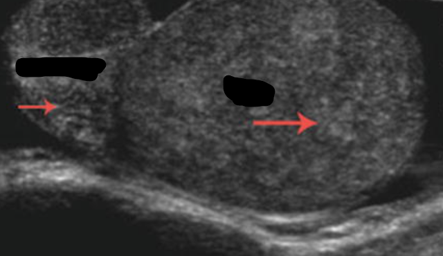
name the finding and give 2 acquired or 2 congenital etiology.
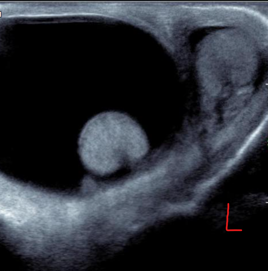
View Answer

name the finding and give 2 acquired or 2 congenital etiology.


Hydrocele

There are two main subtypes of congenital hydrocele:

  • communicating hydrocele

  • spermatic cord hydrocele

In the communicating type, fluid collects around a patent processus vaginalis which failed to successfully obliterate.

The spermatic cord hydrocele is further subdivided into:

  • encysted type (spermatic cord cyst): no communication with the peritoneum or tunica vaginalis

  • funicular type (funiculocele)

    • communicates with the peritoneum at the internal ring and does not surround the testis

    • more common in children and premature infants 

  • trauma

  • epididymitis

  • testicular torsion

  • testicular neoplasm

  • testicular infarction 


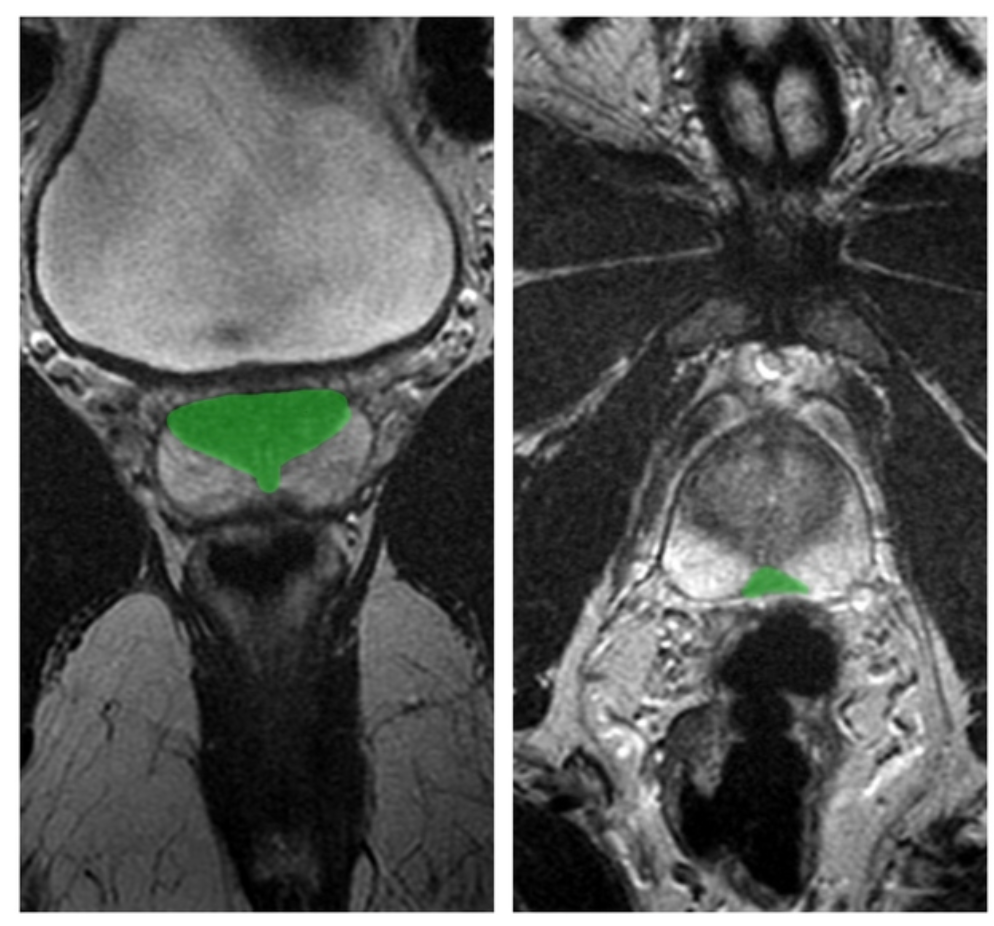
Which zonal prostate anatomy is highlighted in green on these coronal and axial T2 images of the prostate gland?

View Answer

Which zonal prostate anatomy is highlighted in green on these coronal and axial T2 images of the prostate gland?


central zone


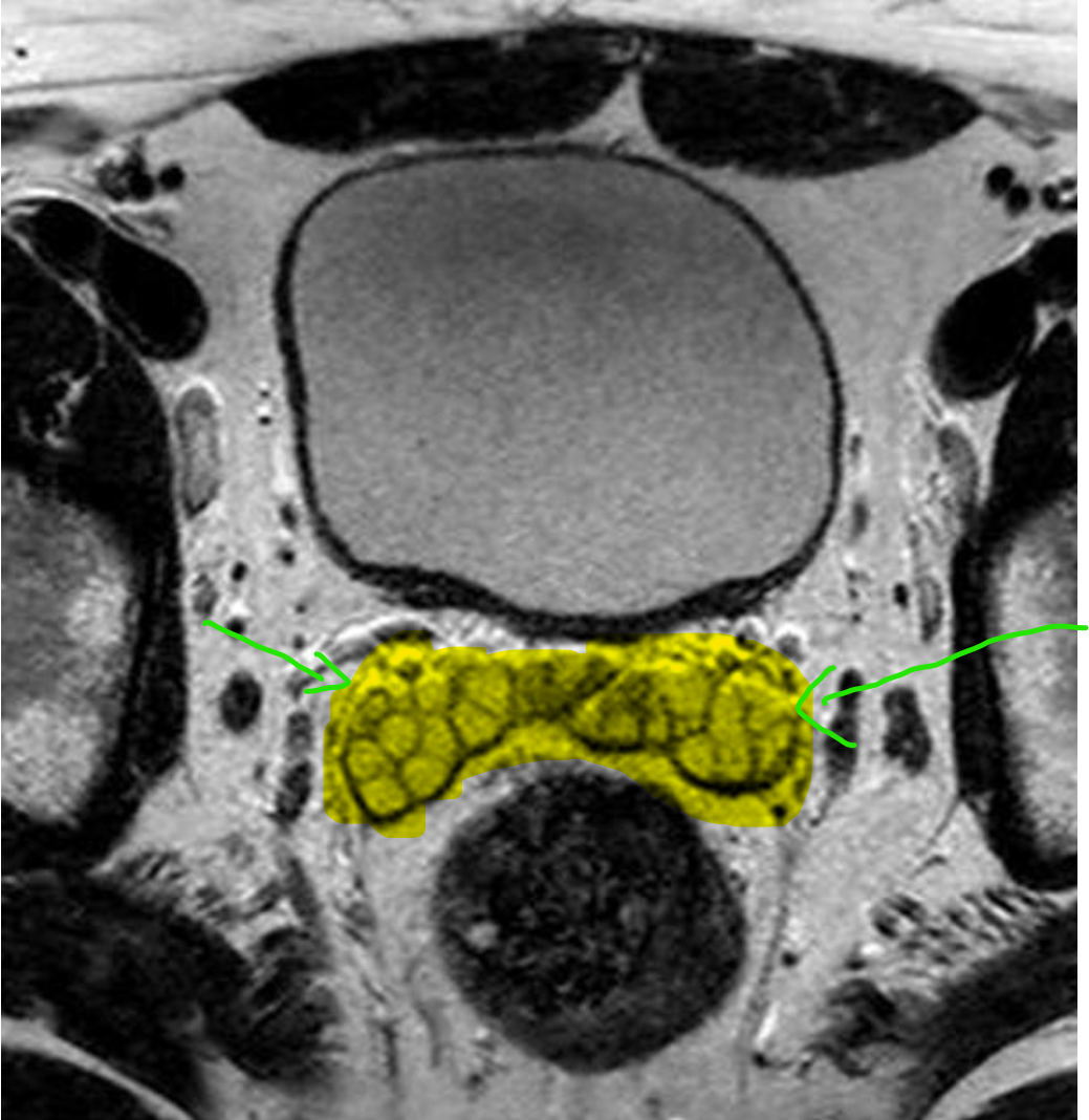
Which anatomical structure is pointed at on these axial T2 images?

View Answer

Which anatomical structure is pointed at on these axial T2 images?


Seminal Vesicles


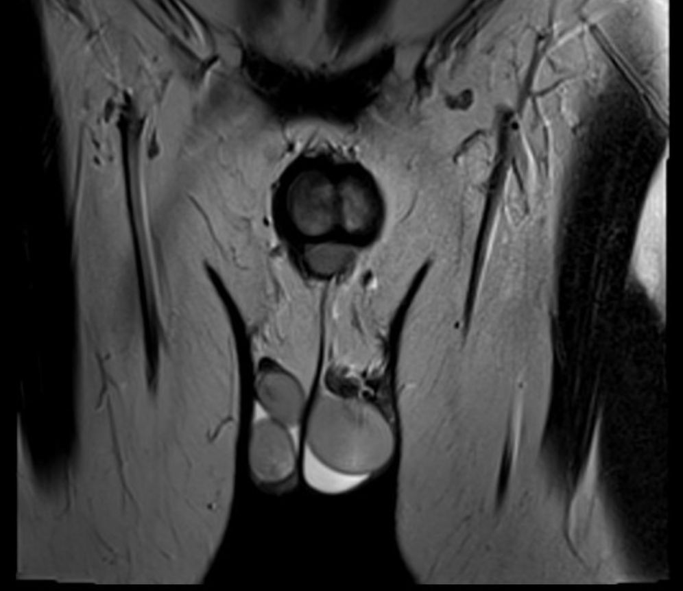
name the finding. is it more common on the right or left?

View Answer

name the finding. is it more common on the right or left?


Polyorchidism / triorchidism

triorchidism, there are three testes with the supernumerary being situated on the left in 65% of the cases.


Jeopardy Event!!!


you can steal 10 points from any team.


if this was the first pick then tough luck. 0_0

View Answer

Jeopardy Event!!!


you can steal 10 points from any team.


if this was the first pick then tough luck. 0_0


if you didn't steal gain 40 points and a hint for final question. void if chosen as first question.


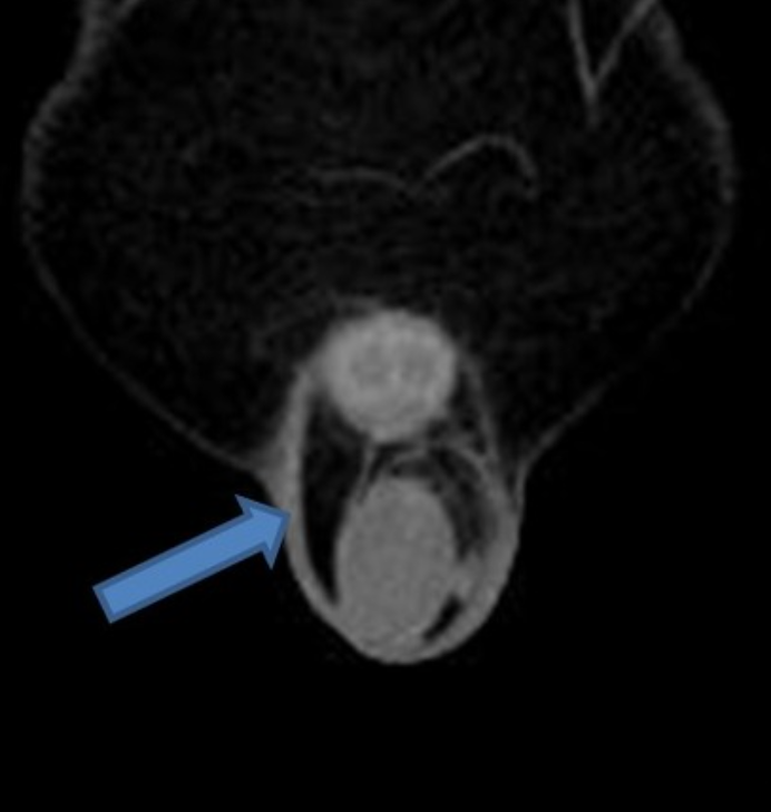
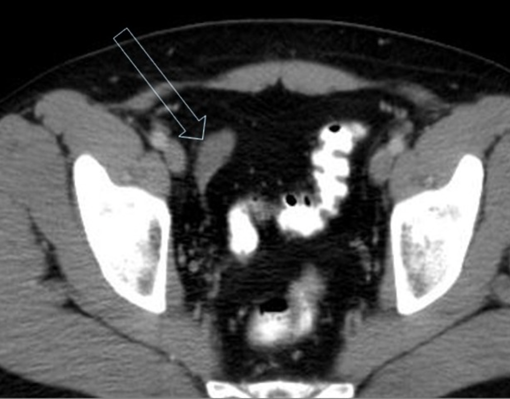
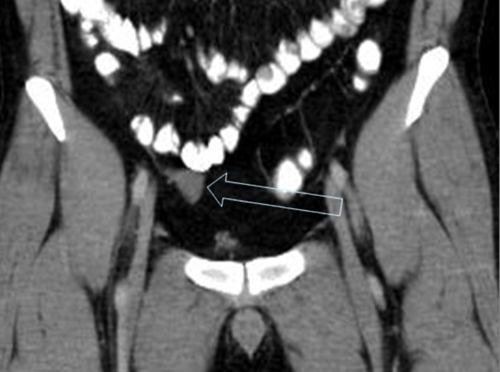
what are the findings?

https://radiopaedia.org/cases/28322/studies/28564?lang=gb#images

View Answer

what are the findings?

https://radiopaedia.org/cases/28322/studies/28564?lang=gb#images


Cryptorchidism 


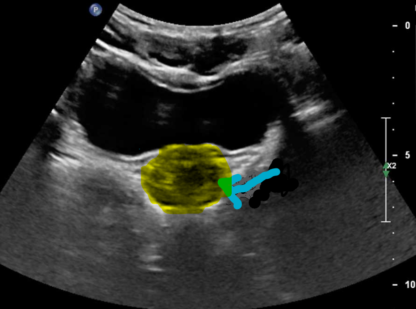
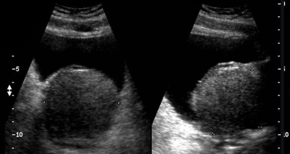
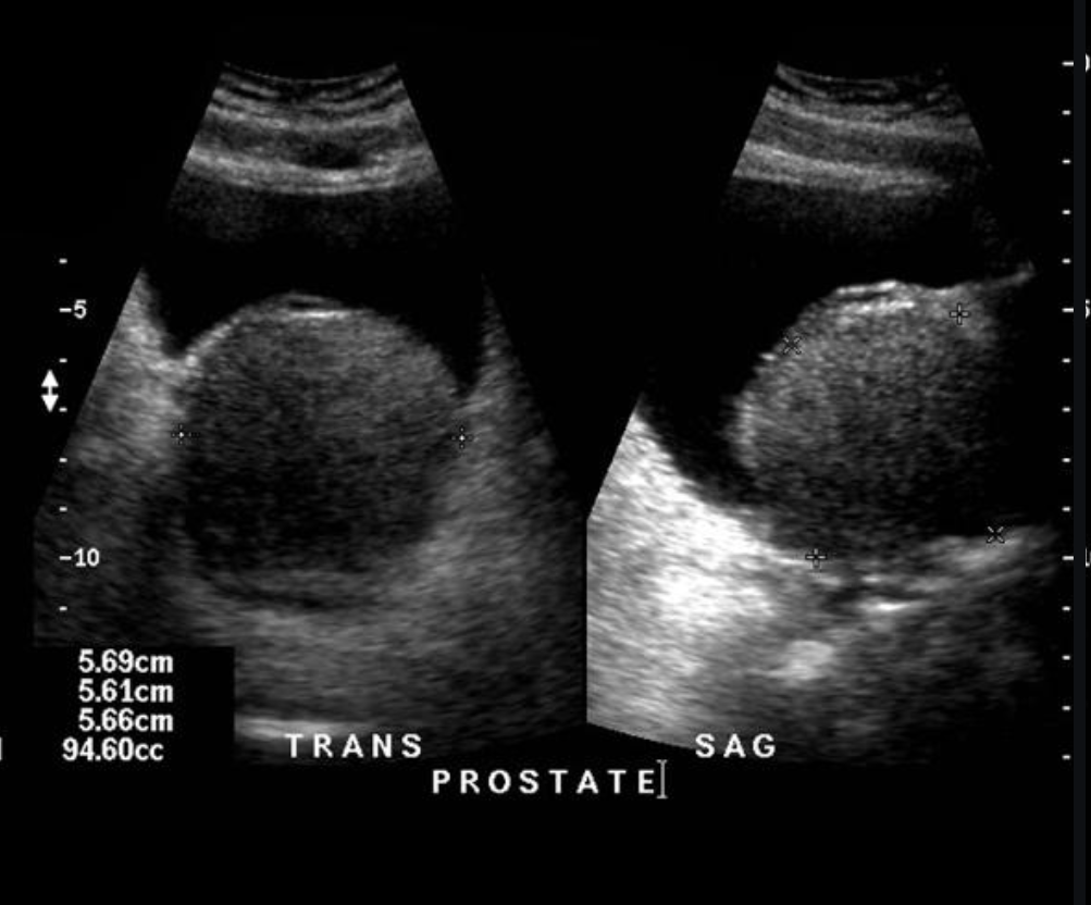
65 Year old male with urinary incontinence and constant bladder fullness post micturition.

what are the findings in this image and the most common diagnosis.

View Answer

65 Year old male with urinary incontinence and constant bladder fullness post micturition.

what are the findings in this image and the most common diagnosis.


Ultrasound of the bladder and prostate demonstrates and enlarged prostate (consistent with benign prostatic hypertrophy)


name 2 central and 2 lateral cystic lesions/cyst-like lesions of the prostate. (you can do 1/3 split)

View Answer

name 2 central and 2 lateral cystic lesions/cyst-like lesions of the prostate. (you can do 1/3 split)


midline cystic lesions/cyst-like lesions

  • Mullerian duct cyst
  • prostatic utricle cyst
  • ejaculatory duct cyst (tends to be more paramedian 6)
  • cystadenoma of prostate
  • ductus deferens cyst
  • TURP defect

lateral cystic lesions

  • cystic degeneration of benign prostatic hyperplasia
  • prostatic retention cyst
  • seminal vesicle cyst
  • diverticular prostatitis
  • prostatic abscess
  • cavitatory prostatitis from chronic prostatitis
  • parasitic prostatic cyst
  • cystic prostatic carcinoma

what is the difference between primary and secondary varicocele?

which testes is more commonly affected and why?

View Answer

what is the difference between primary and secondary varicocele?

which testes is more commonly affected and why?


Most varicoceles are primary and result from incompetent or congenitally absent valves in the testicular vein (internal spermatic vein).

Secondary varicoceles are much less common and result from increased pressure in the testicular vein due to compression (e.g. extrinsic mass such as retroperitoneal lymphadenopathy or renal mass, or renal vein compression in nutcracker syndrome), obstruction (e.g. renal vein thrombus), or splenorenal shunting (portal hypertension). 


The left testis is affected much more commonly affected (≈85%) than the right.


This may be due to the shorter course of the right testicular vein and its oblique insertion into the IVC which creates less backpressure. In contrast, the left testicular vein has a longer course and inserts into the left renal vein at a right angle. 


Bilateral varicoceles are not uncommon (≈15%).


what is the normal testes size?


get 20 extra points if you can tell newborn testes size.

View Answer

what is the normal testes size?


get 20 extra points if you can tell newborn testes size.


Normal adult testes measure approximately 3 cm (AP) x 2-4 cm (TR) x 3-5 cm (length), with a volume of 12.5-19 mL


At birth, testes measure approximately 1.5 cm (length) x 1 cm (width), reaching ~4 mL volume at puberty


what is your primary differential for this case?

https://radiopaedia.org/cases/13513/studies/13445?lang=gb

https://radiopaedia.org/cases/59368/studies/66726?lang=gb#images

View Answer

what is your primary differential for this case?

https://radiopaedia.org/cases/13513/studies/13445?lang=gb

https://radiopaedia.org/cases/59368/studies/66726?lang=gb#images


Prostatic abscess

U/S:

It usually demonstrates ill-defined hypoechoic areas within an enlarged and/or distorted prostate gland. There may be inhomogeneous echoes within these ill-defined areas.


CT:

The prostate is replaced by a large collection of peripherally enhancing, low-attenuation fluid which extends into the left ischioanal fossa. Thickening of the bladder base, likely due to reactive inflammation.


scoring system of the Prostate using MRI is called?

View Answer

scoring system of the Prostate using MRI is called?


PI-RADS (Prostate ImagingReporting and Data System)


when was the first MRI used to test for prostate cancer? (I will give the score with difference of 10)

View Answer

when was the first MRI used to test for prostate cancer? (I will give the score with difference of 10)


1971


first full study published 1982


what is the difference between the 3rd and 4th grade of varicoceles on doppler?

View Answer

what is the difference between the 3rd and 4th grade of varicoceles on doppler?


  • grade III
    • no major dilatation in supine position
    • dilated veins up to lower pole of testis seen only in standing position
    • reflux at lower pole veins during Valsalva manoeuvre


  • grade IV
    • dilated veins even in supine position
    • reflux during Valsalva manoeuvre

Pandora box jackpot!!!!


what is 1 + 1?

View Answer

Pandora box jackpot!!!!


what is 1 + 1?


2


testicular cancer by decade.


fill for 3 decades. or give 4 answers.

  • 1st decade: 2 answers

  • 2nd decade: 1 answers

  • 3rd decade: 1 answers

  • 4th decade: 1 answers

  • ≥7th decade: 2 answers

View Answer

testicular cancer by decade.


fill for 3 decades. or give 4 answers.

  • 1st decade: 2 answers

  • 2nd decade: 1 answers

  • 3rd decade: 1 answers

  • 4th decade: 1 answers

  • ≥7th decade: 2 answers


  • 1st decade: yolk sac tumour and testicular teratoma

  • 2nd decade: choriocarcinoma

  • 3rd decade: embryonal cell carcinoma

  • 4th decade: seminoma

  • ≥7th decade: lymphoma (usually non-Hodgkin lymphoma) and spermatocytic seminoma


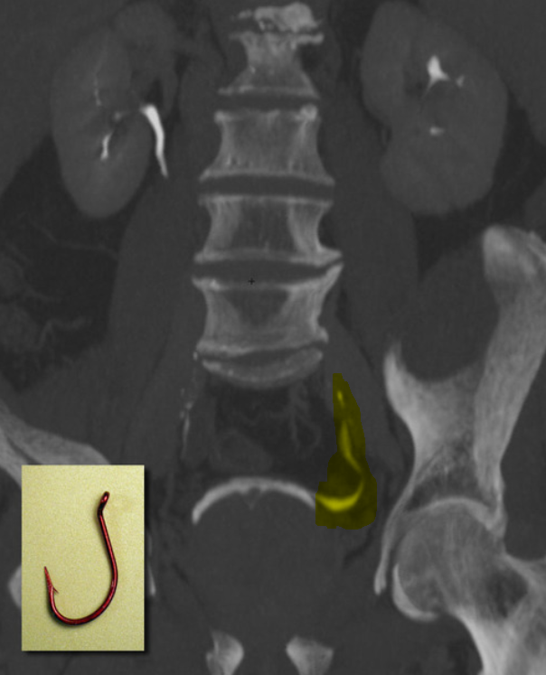
fish hook sign is sign for what?


View Answer

fish hook sign is sign for what?


The fish-hook configuration of the distal ureter indicates bladder outlet obstruction due to benign prostatic hyperplasia.


Scoreboard
Scoreboard
Team 1
Lose Point
Add Point
Team 2
Lose Point
Add Point
Team 3
Lose Point
Add Point
Team 4
Lose Point
Add Point
Team 5
Lose Point
Add Point
Team 6
Lose Point
Add Point
Team 7
Lose Point
Add Point
Team 8
Lose Point
Add Point
Team 9
Lose Point
Add Point
Team 10
Lose Point
Add Point


What Would You Like To Risk?

Scoreboard
Team 1
Minus 1
Plus 1
Team 2
Minus 1
Plus 1
Team 3
Minus 1
Plus 1
Team 4
Minus 1
Plus 1
Team 5
Minus 1
Plus 1
Team 6
Minus 1
Plus 1
Team 7
Minus 1
Plus 1
Team 8
Minus 1
Plus 1
Team 9
Minus 1
Plus 1
Team 10
Minus 1
Plus 1


Go To The Final Question
Final Score:
Scoreboard
Team 1
Lose Point
Add Point
Team 2
Lose Point
Add Point
Team 3
Lose Point
Add Point
Team 4
Lose Point
Add Point
Team 5
Lose Point
Add Point
Team 6
Lose Point
Add Point
Team 7
Lose Point
Add Point
Team 8
Lose Point
Add Point
Team 9
Lose Point
Add Point
Team 10
Lose Point
Add Point


Create a New Game Create a New Game